MRI scans offered at Highland
Published 7:00 am Friday, May 16, 2014
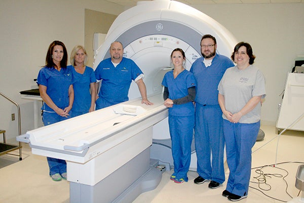
EXPERT IMAGING: Highland Community offers several diagnostic imaging services, including this digital MRI unit.
Photo by Jeremy Pittari
Highland Community Hospital offers a state of the art imaging department with not only x-ray and CT scan capabilities, but also brand new $1 million MRI unit.
James Turnage Jr., Director of Radiology and Cardiology at Highland said the MRI unit at Picayune’s hospital is the latest model offered by GE, offering the first digital magnet.
While it is not the largest MRI unit commercially available, it does offer a larger than average bore, or opening, which helps ease the mind of claustrophobic patients.
An MRI unit, which stands for magnetic resonance imaging, captures images of hard and soft tissues by using magnetic fields to align cells to determine how much they weigh, Turnage said. Each cell in the human body has a predetermined weight, and if that number is off, then an abnormality is detected.
Contrary to popular belief, the unit uses no radiation to capture images.
“When everyone thinks of imaging they always think of radiation,” Turnage said.
While this technology allows medical staff to capture sharp images of soft tissue other methods can’t, there are some slight risks. Turnage said patients with brain stints, metal joint implants or a heart implant will have to have those items crosschecked with the manufacturer to ensure they are MRI safe. This precaution is necessary because the powerful magnet could cause those implants to shift, possibly causing serious injury or death if they are made of certain substances.
“If you get a pace maker you’re never going to have an MRI,” Turnage said.
However, recent advances in medical technology allow for MRI safe implants. The implant’s serial number leads medical staff to manufacturer records that determine if the implant is safe.
“There is no skipping of steps, one skip could mean the end of somebody’s life,” Turnage said.
Another safety precaution includes asking patients to remove all metal objects from their possession, including cell phones.
Patients are asked to empty their pockets not just to ensure their safety, but also to avoid damaging their possessions, such as bank cards that use magnetic strips to store data.
“If you go in there with your wallet you won’t be using your cards anymore,” Turnage said.
Every piece of equipment in the MRI room also has to be safe for use, including stretchers.
To ensure the magnetic fields don’t cause problems for the rest of the facility, the entire MRI room is lined with copper, Turnage said.
These precautions are not only employed to protect the patient, but the equipment as well. If an object is pulled into the magnet, the only way to remove it is to turn the unit off. Shutting the magnet down is an expensive procedure and involves purging the cryogen from the system. Turnage said to refill the system’s cryogen can cost about $150,000, an expense every hospital works to avoid.
There are safety features to ensure the MRI remains functional. The device is networked to the manufacturer who monitors the equipment for potential problems. Turnage said most of the time the manufacturer will know if the equipment needs servicing before a problem occurs and alert the hospital of that information.
While most people would think the image from an MRI unit is captured like a photograph, the data collected by the MRI is actually processed through a computer before an image is shown. This allows software to compensate for a patient’s slight movement and apply special filters as they capture the image to reduce blurry images.
MRI units are capable of capturing images of bone, soft tissue and vessels.
Typically a patient would undergo an MRI scan if they needed to find a ruptured disk in the spine, check the brain for signs of a stroke, locate ligament tears in joints or check their abdomen for signs of cancer, Turnage said.
By utilizing specialized attachments, the equipment is also capable of detecting signs of breast cancer. However, the MRI is used only after less costly methods have been exhausted. Once at that step, using an MRI can prevent the patient from undergoing a biopsy.
Highland is currently working towards receiving their breast imaging center of excellence designation. Turnage said the last requirement is the purchase of stereotactic breast biopsy equipment, which will cost about $160,000. At that point the hospital can apply for the designation.
The time required to capture a typical MRI image varies somewhat, but the average time is about 30 minutes in the unit. Turnage said to scan a skull takes about 20 minutes, while an abdomen scan can take about 45 minutes. The entire process from entering the hospital to checkout is about an hour.
Most patients will not have an MRI scan conducted as a first precaution. Turnage said medical staff will start with an x-ray or CT scan first, not just because they take less time, but because those methods are cheaper for the patient. As a comparison, an x-ray can cost about $200, while and MRI scan will run about $2,000.
An MRI scan is a noisy process. While the model at Highland is quieter than most models, patients are still required to wear earplugs during the procedure.
Turange said to help keep patients entertained during the scan the unit can wirelessly stream music from a device in the other room to their earplugs.
Not every hospital in the nation is equipped with an MRI unit. According to data obtained from the Center for Disease Control in 2007 there were 34.3 MRI equipped facilities per one million people.
“This town and hospital is very fortunate to have an MRI in house,” Turnage said.